Biological-related studies
My research on the mechanics of living cells and their interaction with surfaces began during my postdoctoral work (2000-2002) in the lab of Prof. E. Sackmann in Munich. There, I had the opportunity to study cellular mechanics using magnetic tweezers, exploring how functionalized magnetic beads interact with living cells and soft materials such as hyaluronic acid gels. This work provided fundamental insights into the forces involved in cell adhesion, migration, and deformation. As part of this research, I investigated the protrusion forces generated by phagocytic cells, showing how macrophages use local force transmission to engulf particles [16]. By applying controlled forces to β1 integrins using magnetic tweezers, I demonstrated that forces above a certain threshold induce active formation of protrusions, providing key insights into the early steps of phagocytosis [11]. I later extended this work to amoeboid cell crawling and how these cells transmit forces when moving on soft biological tissues, in two projects managed by Guy Ladam [12, 13].
Building on my expertise in cellular mechanics, I later investigated the behavior of macrophages interacting with nanoparticle-based coatings. I demonstrated that the size of nanoparticles strongly influences their internalization efficiency, highlighting the fragility of such coatings when exposed to cellular uptake [41].
More recently, I have focused on the role of trapped air in bacterial adhesion, particularly in superhydrophobic surfaces. I led a study on how trapped air influences the attachment of Staphylococcus aureus to superhydrophobic elastomer surfaces textured by femtosecond laser [39]. Additionally, I have collaborated on research examining the interaction between Escherichia coli and textured surfaces, focusing on how bacterial colonization is influenced by wetting properties [40].
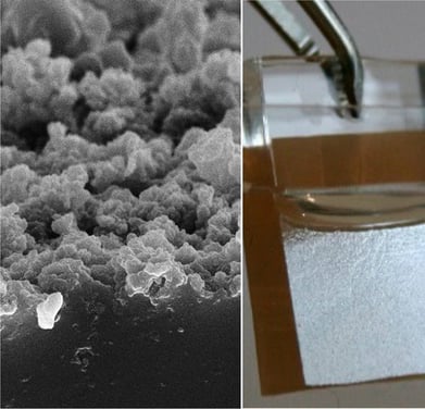
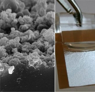
SEM image of a femto-laser textured PDMS surface (left) and air plastron produced in water over this textured surface (right) [39].
Confocal fluorescence microscopy sequence of macrophages internalizing 500 nm green fluorescent particles from a monalyer (total length is about 10 hours) - Tatiana Petithory [41]